
 EmbryoScope is an embryo-selection tool. This high-tech device is used in the IVF laboratory during the embryo culture period between fertilisation and implantation.
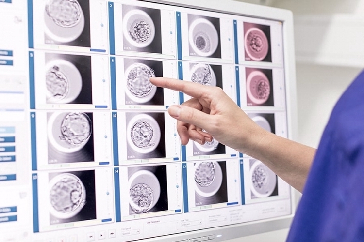
EmbryoScope uses time-lapse imaging, resulting in the selection of the best embryo for transfer, ranking the entire development process and enabling the prediction of the embryo with the best viability.
MONITORING EMBRYOS WHILE THEY GROW
The EmbryoScope captures information about developing embryos in its special incubator. An incubator maintains the necessary physiological conditions required by a living embryo while they are in the IVF laboratory. A high-resolution time-lapse camera and a computer with special software monitor entire embryo development.
Incorporated time lapse system continuously captures images and records them as a video of the embryonic development, allowing the embryologist to monitor embryo cell divisions and carefully observe its development without any need to manipulate with embryos while cultivating.
Being able to watch the embryos grow without exposing them to conditions outside the incubator is one of the many benefits. EmbryoScope also uses learning algorithms constantly searching the data for patterns, and growing evidence suggests that there are certain key time points in development that are tied to positive or negative results.
EmbryoScope helps the embryologist predicting the further development and pregnancy potential of each embryo. These crucial developmental changes can only be observed with the help of the EmbryoScope’s time lapse camera and the very best embryos can be selected for transfer.
EMBRYOS GROW IN AN UNDISTURBED ENVIRONMENT
The embryo transfer usually takes place 5-6 days after the fertilisation. But not all embryos are capable of leading to a pregnancy. Embryos vary in quality. The better the quality, the more likely an embryo is to implant and lead to a successful pregnancy. EmbryoScope assists with selecting the embryo with the highest chance.
Observing features of an early embryo development is essential for evaluation of the embryo’s potential to implant and become a successful pregnancy. The traditional ‘snap-shot’ evaluations may miss critical embryo development patterns.
THE TRADITIONAL WAY OF EMBRYO SELECTION
With or without EmbryoScope, collected eggs and sperm are combined in a sterile dish filled with a special culture medium for growth. Than the embryos need to be cultured in an incubator, which copies the conditions inside a woman’s body.
Without using the EmbryoScope embryologist must remove the embryo from the mini incubator to perform three to five brief evaluations of the developing under the microscope.
The evaluation time allowed for these ‘snap-shots’ is limited by the need to minimise the time embryo spends outside the safe environment of the incubator. This is to avoid embryo stress which can reduce the quality of the embryo and therefore the chances of pregnancy.
Traditional evaluation is likely to miss abnormal cleavage patterns. One abnormal cleavage pattern is direct cleavage which has been shown to occur in up to 26% of embryos and to reduce chances of implantation after transfer. This is when embryo divides directly from 1 to 3 cells instead of dividing normally from 1 to 2 to 4 to 8. Another abnormal cleavage pattern is when embryo goes from a higher to a lower cell number.
DOES EMBRYOSCOPE WORK?
The cell division pattern of the embryos is becoming a valid alternative for selecting the embryos which have the best potential for implantation. There is growing evidence suggesting that certain cell time points and events are especially important for predicting further development potential, and pregnancy potential of embryos.
Some retrospective studies have shown that embryos with specific division times and certain development patterns can have up to 15 – 20% better chance of pregnancy. The optimum times for cell division can be checked more easily and the chances of implantation improved in cases in which selection using EmbryoScope technology is possible.
WHO IS EMBRYOSCOPE SUITABLE FOR?
Although in theory this technology can be applied to any type of patient undergoing IVF treatment the greatest improvement is among patients who repeatedly produce multiple abnormally developing embryos.
It can be used in cases where more information about the embryo is desired in situations where there is repeated implantation failure, advanced maternal age and history of recurrent miscarriage. It will help couples and women to make an informed decision about future treatment plan or closure as appropriate.
ARE THERE ANY RISK LINKED TO EMBRYOSCOPE USAGE?
There are no risks involved in using time-lapse imaging to monitor developing embryos. What is important to realise, and is only logical, that because the EmbryoScope gives embryologist such highly-detailed information, it may result that the information gathered show that there are fewer or no embryos suitable for transfer or freezing.
ADVANTAGES OF THE EMBRYOSCOPE
The Embryoscope allows more frequent imaging, therefore, more information about the crucial early developments. It helps experts identify potential valuable information about the embryo and its potential to survive transfer and implant, all while keeping the embryo in the best culture condition. Anomalies can be shown in EmbryoScope that may not be visible during traditional embryo culture.
Keeping the embryo in this controlled and protected environment at all times helps to eliminate risks involved in moving and examining developing embryos but allowing all the potential for the selection of the best embryo for transfer.
__ __
TIP: what to read on our blog next: Are you familiar with the IVF procedure? Find out: IVF treatment: Basic recap overview